
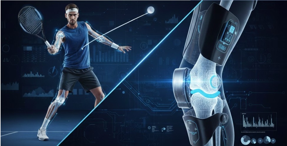
AI-Driven 3D Reconstruction of Knee anatomy from MRI: Bridging Functional Assessment and Precise Anatomical Modeling
Knee injuries, particularly anterior cruciate ligament (ACL) tears, and degenerative conditions such as osteoarthritis represent major burdens on global healthcare systems. Accurate 3D reconstruction of knee anatomy from MRI data, combined with real-time functional assessment via wearable exoskeletons, offers a powerful pathway toward personalized diagnosis, preoperative planning, and rehabilitation. This article reviews recent breakthroughs in two complementary domains: (1) modular active knee exoskeletons equipped with multi-modal sensing (IMU, force, EMG) for dynamic biomechanical data collection, and (2) self-supervised deep learning models, such as Vision Transformers (ViT) pretrained with masked autoencoding and BYOL-based approaches, for high-fidelity 3D knee reconstruction from 2D MRI slices. We highlight our prototype exoskeleton and AI pipeline developed at the Center for Excellence in Medical Robotics and Rehabilitation (CEMRR) at Nazarbayev University, Astana, demonstrating how these technologies can be integrated to correlate structural pathology with functional deficits, ultimately enabling more effective, patient-specific interventions.

The Clinical Need: From Static Images to Dynamic Function Magnetic resonance imaging (MRI) remains the gold standard for visualizing knee joint pathology, yet conventional 2D slice-by-slice analysis often fails to capture the complex 3D relationships among bones, cartilage, menisci, and ligaments. Three-dimensional reconstructions provide a holistic view essential for accurate diagnosis, surgical planning, and postoperative monitoring. However, static anatomical models alone do not reveal how injuries affect movement, loading, or muscle activation patterns.
Active knee exoskeletons address this gap by delivering controlled mechanical support while simultaneously collecting rich kinematic, kinetic, and neuromuscular data during real-world tasks. Over the past two decades, the field has progressed from passive spring-damper systems to powered devices capable of generating 30–60 N·m torques. Modern prototypes increasingly adopt modular, 3D-printed designs and advanced control strategies (impedance/admittance, assist-as-needed, EMG-driven intention recognition) to enhance biomechanical compatibility and user transparency.
Our Modular Active Knee Exoskeleton Prototype

At CEMRR, we have designed and fabricated a lightweight, modular rigid-frame knee exoskeleton optimized for clinical research and rehabilitation (Figs. 20–25). Key features include:
Lateral drive unit - An electric motor with high-ratio gearbox positioned close to the knee joint to minimize inertia and moment arms.
Single-axis motorized hinge - Allows bidirectional flexion/extension with low-resistance passive mode when unpowered for safety.
3D-printed modular architecture - Femur and tibia segments are easily customizable for different anthropometries and enable rapid swapping of motors or sensors.
Multi-modal sensing suite:
IMUs on thigh and shank for real-time knee angle, angular velocity, and orientation.
Insole force/pressure sensors for ground-reaction-force timing and gait symmetry.
Planned integration of surface EMG to capture quadriceps/hamstrings activation and load asymmetries.
Pilot data collection in healthy volunteers (target N ≥ 30) has already yielded time-series profiles of normative movement patterns. These dynamic measurements complement static MRI, enabling clinicians to link imaging findings (e.g., meniscal tears, ligament damage) to functional limitations such as restricted flexion arcs, reduced peak torque, or asymmetric weight-bearing.
AI-Powered 3D Reconstruction: Self-Supervised Learning Breakthroughs
To overcome the labor-intensive nature of manual 3D segmentation from 2D MRI, CEMRR has developed and validated self-supervised deep learning pipelines that require minimal labeled data.
Vision Transformer with Masked Autoencoding (MAE)
MRI slices are divided into fixed-size patches, projected into embeddings, and processed by a ViT encoder with multi-head self-attention. During pretraining, random patches are masked; a lightweight decoder reconstructs them, forcing the model to learn robust anatomical representations. A subsequent 3D convolutional decoder aggregates multiplanar information (axial, sagittal, coronal) to produce coherent volumetric models. Training curves show stable convergence with strong Dice, IoU, and Recall scores. STL-exported reconstructions preserve bone geometry, proportions, and articular surfaces with high fidelity to manual segmentations in 3D Slicer.
BYOL-Based ACL Tear Detection and Segmentation
A parallel effort employs Bootstrap Your Own Latent (BYOL) self-supervised pretraining followed by a U-Net-like architecture for precise segmentation of ACL tears. This approach significantly outperforms standard supervised U-Net models when labeled data are scarce, as demonstrated in recent work published in MethodsX (doi:10.1016/j.mex.2025.103664). Visual comparisons confirm superior boundary delineation and reduced false positives.
Integration: Toward a Unified Diagnostic and Rehabilitative Pipeline
The true innovation lies in combining these technologies. Exoskeleton-derived functional data (joint angles, torques, muscle activations) can be mapped directly onto AI-reconstructed 3D anatomical models. This fusion enables:
Personalized rehabilitation. Assist-as-needed torque profiles tailored to the patient’s specific injury pattern and recovery trajectory.
Preoperative planning.Surgeons visualize both structure and predicted function.
Longitudinal monitoring. Track biomechanical changes post-surgery or during conservative treatment.
Broader Impact at CEMRR
These efforts are part of CEMRR’s broader mission to advance AI in medicine and rehabilitation. Complementary AI projects include radiomics for orthopedic and oncologic research, as well as sports-performance systems (e.g., AI-PIONEER for elite butterfly swimming evaluation in collaboration with the National Olympic Council of Kazakhstan, and FAST-AI for tennis serve analysis). Such interdisciplinary work positions Kazakhstan as an emerging hub for intelligent medical and sports technologies.
Looking Ahead
Challenges remain, actuator mass, battery life, standardized benchmarking, and regulatory pathways for clinical adoption. Yet the rapid convergence of wearable robotics and self-supervised AI heralds a new era of precision orthopedics. Future work at CEMRR will focus on full clinical validation, soft-tissue reconstruction, and seamless integration into hospital workflows.
